Wednesday, November 30, 2022

Meckel's Scan


  • Most common congenital anomaly of GI tract (INCIDENCE – 1 to 3% of general population).
  • Most common cause of lower gastrointestinal haemorrhage in previously healthy infants.
  • Vestigial remnant of the omphalomesenteric duct.


  • Location – Anti-mesenteric border of the terminal ileum within 80–100 cm of the ileocecal valve. 
  •  Approximately 57% of Meckel diverticula contain heterotopic gastric mucosa -secretes HCL.
  • Most common sign of Meckel diverticulum is gross rectal bleeding.
  • 99mTc-pertechnetate is taken up by the mucin-producing cells of gastric mucosa and is then secreted into the gut lumen.
  • Overall sensitivity of 85%, specificity of 95%, and accuracy of 90%. 

  • CLINICAL INDICATIONS:
  • To localize heterotopic gastric mucosa in a Meckel diverticulum as the source of unexplained GI bleeding.
  • When patient is not actively bleeding – Meckel scintigraphy.
  • Active bleeding -radiolabelled red blood cell (RBC) scintigraphy.

  • PROCEDURE:
  • Rule out recent in vivo RBC labelling, recent barium studies, colonoscopy and use of laxatives.
  • Fasting of 3–4 h.
  • Ensure voiding before study and and at the end before imaging.
  • Histamine H2 blockers, Proton pump inhibitors, Glucagon (TO IMPROVE SENSITIVITY)
  • 99mTc-pertechnetate is injected IV.
  • Recommended dose
  •               Children - 0.05 mCi/kg  (minimum – 0.25 mCi)
  •               Adults - 8–12 mCi

  • ACQUISION PROTOCOL:
  • Supine position
  • Imaging field – abdomen and pelvis (to include stomach and bladder)
  • Children – thorax should be included
  • Anterior abdominal dynamic flow images
  • 1–5 s/frame for up to 1min, 30-60s per frame for 30-60 min
  • Static images (anterior, anterior oblique, lateral, and posterior projection views) are recommended at the end of the dynamic acquisition.
  • SPECT CT may improve the detection of 
  •                     - small diverticulum.
  •                     - diverticulum obscured by the urinary bladder.
  •                     -clinical suspicion for a Meckel diverticulum is high and the planar images have negative or equivocal findings.

  • MECKEL'S SCAN


  • Normal uptake in flow phase – heart, lung, major vessels, liver, spleen, kidney, ureter and bladder.
  • Stomach activity appears early on dynamic scintigraphy and is most prominent after 10–15 min.
  • Ectopic gastric mucosa is visible as a focal, localized area of uptake that appears at the same time as the activity in the normal gastric mucosa.
  • Most common location – right lower quadrant.
  • A four year old child presented with recurrent episodes of pain abdomen around the umbilicus associated with passage of fresh blood per rectum off and on for the last one and half years.
  • Prominent gastric activity appearing at approximately 15 min in the left upper quadrant.
  • Small round area of intense tracer activity in the right lower quadrant of abdomen suggesting the presence of ectopic gastric mucosa.
  • Exploratory laparotomy which confirmed the presence of Meckel’s diverticulum.
  • Diverticulectomy with end to end anastomosis.


  • FALSE POSITIVE:
  • Tracer activity in the proximal small bowel, kidneys, ureter, or bladder is mistaken for ectopic gastric mucosa.
  • Duplication cyst with ectopic gastric mucosa
  • Bowel inflammation 
  • Intussusception or small-bowel obstruction 
  • Peptic ulcer 
  • Vascular lesions with increased blood pool (hemangioma or AV malformation)

  • FALSE NEGATIVE:
  • 1. Procedures that may cause interference
  •                     - barium fluoroscopy, administration of perchlorate.
  • 2. Anatomic or physiologic causes of errors – when image is obscured by
  •                     - brisk GI bleeding
  •                     - by the urinary bladder or dilated ureter
  •                     - focus of ectopic mucosa is small (1.8 cm 2)
  •                     - if there is movement of the diverticulum


Follow me on Twitter @RutujaKote for more discussions about topics in Nuclear Medicine and Radiology.

Tuesday, November 29, 2022

MUGA


  • Multigated acquisition (MUGA)

  • Equilibrium radionuclide angiocardiography (ERNA)

  • Radionuclide ventriculography (RVG)


  • Procedure in which patient’s RBCs are radiolabeled and gated cardiac scintigraphy is obtained.

  • Data are collected from several hundred cardiac cycles to generate an image set of the beating heart that is presented as a single, composite cardiac cycle.


Monday, November 28, 2022

Langerhans Cell Histiocytosis


  • Histiocytic disorders are a group of diseases derived from macrophages and dendritic cells, which are important antigen presenting cells and mediators of host immune responses
  • Dendritic cell disorders consists of:
  1. Langerhans cell histiocytosis (LCH) – Incidence 4 to 4.5 per million
  2. Erdheim-Chester disease (ECD)
  3. Juvenile xanthogranuloma (JXG)
  • Macrophage related disorders include:
  1. Rosai-Dorfman disease (RDD)
  2. Hemophagocytic lymphohistiocytosis (HLH)

Langerhans Cell Histiocytosis:

  • LCH is the most common dendritic cell disorder
  • Caused by abnormal monoclonal proliferation and accumulation of pathological Langerhans cells in tissues.
  • These histiocytes can infiltrate any organ, therefore the clinical presentation and prognosis are highly variable.
  • Unique intracytoplasmic organelles, known as Birbeck granules which appear racquet shaped in electron microscopy helps to differentiate LCH from other disorders.
Types:

  • According to number and types of organs involved, LCH is classified into:
  • Single system LCH – which involves only one organ or system, particularly bone. It can be:
  1. Unifocal/localized seen in approx. 70% of cases, usually present between 5 and 15 years of age
  2. Multifocal (chronic recurring form) seen in approx. 20% of cases, presents between 1 and 5 years of age


  • Multisystem LCH – which affects  ≥ 2 organs or systems, seen in 10% of cases, usually diagnosed in first 2 years of life

                   Multisystem LCH is subdivided into low-risk and high-risk groups; high-risk group shows involvement of organs such as liver, spleen, lung, hematopoetic system and CNS, leading to less favorable prognosis.


Investigations:

  • Conventional imaging modalities included radiographs, skeletal surveys, bone scan, CT, MRI and ultrasound
  • Advantages of PET/CT:
  1. Identifies metabolically active histiocytes along with anatomical localization in a single study
  2. Has the potential to detect new lesions that are difficult to diagnose clinically by physical examination and conventional imaging
  3. Helps in monitoring response to therapy.
  • Disadvantages:

  1. Less useful in evaluating lung disease due to variable FDG uptake depending on morphological pattern
  2. Evaluating intracerebral LCH lesions like the pituitary stalk is difficult due to adjacent cerebral cortical uptake.

Bone lesions:

  • Bone lesions occur in approx. 80% of patients
  • Predilection for flat bones – skull is m/c involved, followed by mandible, ribs, pelvis and spine.
  • Skull lesions may be asymptomatic or present with focal pain and scalp swelling.

  • Well-defined lytic punched out appearance of skull lesions is due to asymmetric destruction of inner and outer cortices, which results in characteristic beveled edge
  • When temporal bone is affected, classic location is mastoid and patients present with swelling, dizziness, vertigo and otorrhea
  • Unlike other osseous lesions in LCH, calvarial disease lacks periosteal reaction
  • Skull lesions may grow in size and coalesce, creating a map-like appearance referred to as geographic skull.
  • Vertebral body is the m/c affected part of the spine in LCH
  • Early lesions appear lytic and as disease progresses it can result in symmetric uniform collapse of the vertebral body termed vertebra plana
  • May result in pain and substantial neurological deficits
  • RT is Rx of choice for vertebral lesions at risk for collapse
  • Spinal fusion or bracing may be required in pts with neurological symptoms.
  • Long bone involvement is more common in children than in adults
  • May be asymptomatic or present with focal pain and swelling and are at risk for pathological fractures
  • Femur, humerus and tibia are most commonly involved long bones and lesions typically occur in diaphysis or metaphysis
  • Early lesions appear lytic, expansile and aggressive
  • Less aggressive lesions show cortical thickening and a smooth periosteal reaction
  • Bone scan typically shows increased tracer uptake; however, false negatives are common, particularly with older lesions. Bone scan may also remain positive for a long period for fully treated lesions
  • As lesions become chronic, they may resolve or appear sclerotic from periosteal new bone formation.
  • Whereas long bone lesions are more frequently encountered in children, isolated flat bone involvement (most commonly rib or mandibular lesions) is more frequent in adults
  • Severe mandibular lesions may destroy the alveolar ridge, resulting in a radiographic appearance of “floating teeth” – patients may present with gingival bleeding and facial swelling
Treatment of Bone lesions:
  • Rx of a single skull lesion  includes curettage, curettage with intralesional steroid injection or radiotherapy
  • Chemotherapy and oral systemic steroids may be used to Rx multiple skull lesions or to prevent CNS extension if the mastoid, temporal or orbital bones are involved.
Liver, Spleen and Lymph node involvement:
  • Liver is involved in approx. 15% of cases, findings include hepatomegaly, focal solid or cystic lesions and hepatic dysfunction
  • A potential complication is sclerosing cholangitis, which is progressive and may eventually require liver transplantation.
  • Spleen is involved less frequently than liver. Splenomegaly can lead to cytopenias secondary to hypersplenism
  • Lymph nodes are involved in approx. 20% of cases and most commonly in neck, manifesting as hard or soft matted groups of nodes with lymphedema
  • Rx includes chemotherapy and steroids.
CNS Involvement:
  • CNS is involved in approx. 16% of cases
  • If facial bones or anterior and middle cranial fossa are affected, incidence of CNS involvement is higher, affecting up to 25% of patients
  • m/c clinical manifestation is diabetes insipidus, secondary to infiltration of posterior pituitary gland
  • Growth hormone deficiency has been recognized as occurring later in life in pts with LCH
  • Pts may also present with ataxia and cognitive dysfunction from neurodegeneration, when lesions occur in cerebellum and basal ganglia
  • Rx includes chemotherapy with or without steroids in cases of active disease with diabetes insipidus. Retinoic acid and intravenous immunoglobulins may be added if there are neurodegenerative symptoms.
Lung Involvement:

  • Lung involvement occurs in approx. 10% of cases
  • More common in adults and is almost always associated with smoking
  • Pts usually present with shortness of breath and non-productive cough and sometimes with fever or weight loss
  • In CT, LCH is characterized by  centrilobular micronodules with a predominantly bilateral symmetrical upper-to-mid-lung distribution, costophrenic angles are usually spared
  • As disease progresses, cysts develop and can eventually become the predominant finding
  • Cysts vary in size, usually less than 1 cm. A confluence of cysts may result in a bullous formation, which predisposes to spontaneous pneumothorax
  • PET/CT – Nodular disease appears more likely to yield a positive PET/CT, while cystic disease does not exhibit FDG uptake. Also, size of lesion could be related to metabolic behavior due to resolution power and partial volume effect.
  • Rx includes smoking cessation as well as chemotherapy and steroids in persistent or progressive cases.
Follow me on https://twitter.com/KoteRutuja?t=Tu07QLiziJCR_Qf8NyibZw&s=08 to learn more about important articles, topics and cases in Radiology and Nuclear Medicine.

Saturday, November 26, 2022

Extravasation of Contrast Media

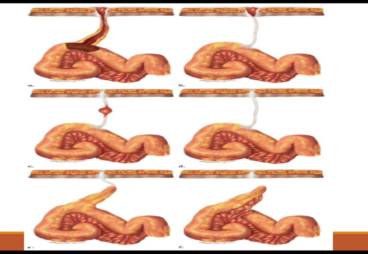
 The reported incidence of intravenous (IV) contrast media extravasation in adults and children ranges from 0.1% to 1.2%.

  • Can also occur with hand injection.

  • may occur at both low and high flow rates.

Complains:

Patients complain of initial swelling or tightness, and/or stinging or burning pain at the site of extravasation, some experience little or no discomfort. On physical examination, the extravasation site may be edematous, erythematous, and tender.

  • In most patients, initial swelling and tenderness resolves within hours to days after the extravasation. The vast majority of patients in whom extravasations occur recover without clinically important sequelae.

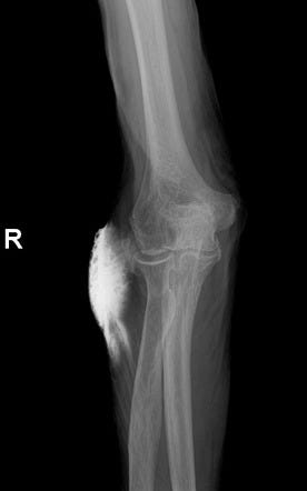
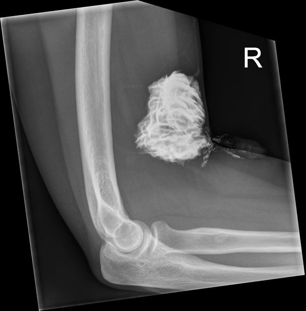
  • However, in some patients, extravasated iodinated contrast media can result in injury to surrounding tissues, particularly the skin, producing an acute local inflammatory response that peaks at 24 to 48 hours. Most of the time there are no lasting complications. 

  • Only rarely will a low-osmolality contrast media (LOCM) extravasation injury proceed to a severe adverse event. Acute tissue injury resulting from extravasation of iodinated contrast media is probably related at least in part to its hyperosmolality.

Severe Complications:

  • The most commonly reported severe injury after LOCM extravasation is compartment syndrome

  • . Compartment syndrome results from mechanical compression and is probably more likely to occur after extravasation of larger volumes of contrast media; however, it also has been observed after extravasation of small volumes, especially when these occur in less capacious areas (such as over the ventral or dorsal surfaces of the wrist).

  • Compartment syndrome may develop soon after an extravasation or result from swelling that sometimes occurs hours after the extravasation.

  • Less commonly encountered severe injuries include skin ulceration and tissue necrosis. These can occur within hours or days of the extravasation event.

Evaluation:

  • Ask about symptoms of pain and paresthesias. 

  • A brief examination should be performed and should include assessment of extremity tenderness, swelling, erythema, paresthesia, active and passive range of motion of the fingers, and perfusion.

Management:

  • Extravasations that resolve rapidly probably need no treatment other than brief observation and discharge instructions.

  • More symptomatic extravasations may be treated with extremity elevation above the level of the heart (to decrease capillary hydrostatic pressure and thereby promote resorption of extravasated fluid and either warm or cold compresses that an outpatient may continue intermittently at home until symptoms resolve, along with discharge instructions, or may be continued on the wards for inpatients.

  • Aspiration is not recommended.

  • Topical application of silver sulfadiazine ointment and steroid cream three to four times daily has been recommended by some as an approach to soothe irritated skin, reduce inflammation, and to prevent infection should any blistering occur, although the efficacy of this treatment is unknown.

  • There is no consistent evidence that local injection of potentially therapeutic agents, such as corticosteroids or hyalurinidase is beneficial.

Surgical Consultation:

  • Urgent surgical consultation should be obtained whenever there is concern for a severe extravasation injury. 

  • Although consultation can prolong length of stay, it should be obtained for any patient in whom one or more of the following extravasation-related signs or symptoms develops: severe pain; progressive swelling or pain; altered tissue perfusion as evidenced by decreased capillary refill; change in sensation in the affected limb; worsening passive or active range of motion; and skin ulceration or blistering.

  • Because of this, surgical consultation should be based on signs and symptoms rather than an absolute volume threshold. If the patient is asymptomatic or has only mild symptoms, appropriate evaluation and clinical follow-up are usually sufficient.

Discharge the patients:

  • Outpatients who have suffered contrast media extravasation should be released from the radiology department only after an initial period of observation, provided the radiologist is satisfied that any signs and symptoms that were present initially have improved or that new symptoms have not developed during the observation period. 

  • Clear instructions should be given to the patient to seek additional medical care for severe pain, progressive pain, numbness or tingling, diminished range of motion (active or passive), skin ulceration, or other neurologic or circulatory symptoms.

  • This is because initial symptoms of a serious compartment syndrome may be absent or relatively mild (such as limited to the development of focal paresthesia).

Patients who are at increased risk of extravasation:

  • Extravasations are more common in patients who cannot communicate effectively (e.g., the elderly, infants and children, and patients with altered consciousness), severely ill or debilitated patients, and patients with abnormal circulation in the limb to be injected. 

  • Patients with altered circulation include those with atherosclerotic peripheral vascular disease, diabetic vascular disease, Raynaud’s disease, venous thrombosis or insufficiency, or prior radiation therapy or extensive surgery (e.g., axillary lymph node dissection or saphenous vein graft harvesting) in the limb to be injected. 

  • Women may have a mild increased risk of extravasation. 

  • Some of these conditions are systemic and cannot be avoided by choosing a different injection site.

  • Certain intravenous access sites (e.g., hand, wrist, foot, and ankle) are more likely to result in extravasation and should be avoided, when possible. 

  • However, use of these alternate injection sites may be necessary due to lack of availability of the more traditional locations. 

  • Flow rates of up to 3 and 5 mL/sec can be safely achieved through 22 gauge and 20 gauge intravenous catheters, respectively, in the vast majority of patients, provided that there is no increased resistance or pain during a rapid test injection. 

  • Patients injected with more viscous contrast material may be more likely to have extravasations than are patients injected with less viscous contrast material. This effect may be mitigated for viscous media by extrinsic warming to human body temperature prior to injection. 

Prevention:

  • Methods for reducing the risk of extravasation include: meticulous intravenous line insertion technique, using angiocatheters instead of butterfly catheters, confirming position by aspirating blood (although failure to aspirate blood does not exclude the possibility of proper catheter location), flushing an inserted catheter with a test injection of saline to ensure proper flow into the accessed vein, and carefully securing the inserted catheter. 

  • Low-risk intravenous line insertion sites are preferred when feasible. If not feasible, higher risk sites may be considered depending on the risks and benefits of administrating contrast media for the examination indication. 

  • Use of a preliminary saline flush to assess injection pressure prior to contrast media administration has been advocated by a few investigators; however, this has not been adopted by most institutions. 

  • Some have recommended use of a hand-held alarm, which the patient can press should any new symptoms develop. 

  • Interestingly, the practice quality improvement project created by the ACR to assist practices in identifying improvements that could be made to reduce the frequency of extravasations did not find that any significant improvement could be achieved, even when risk factors for extravasations were identified and attempts were made to reduce the extravasation risk in advance.


Monday, November 21, 2022

PERCIST 1.0


  • PET Response Criteria In Solid Tumours 

    When to perform?

    • After the first cycle just before the next one

                 -Detect primary resistance to chemotherapy

    • 10 days after the last cycle / End-of-Treatment : 

                 -Evaluates restaging

                 -If resistance developed mid way

    Required parameter for follow up PET-CT study :

    • Fasting for 4-6 hrs

    • Fasting BG < 200mg/dl

    • 50-70 min post injection

    • FU Scan within 15 min of the baseline

    • Same scanner

    • </= 20% activity

    • To be assessable, difference b/w baseline and follow up SUL in liver must be less than or equal to 20%

      SUL peak:

      The SUL peak (this is a sphere with a diameter of approximately 1.2 cm—to produce a 1-cm3-volume spheric ROI) centered around the hottest point in the tumour foci should be determined, and the image planes and coordinates should be noted (SUL peak).

          Background Liver Activity:

    • 3cm ROI mid way between dome & inferior margin excluding central ducts & vessels

    • If diseased –mediastinal background taken, cylindrical ROI in descending aorta, 1cm diameter & 2m long axis.


      Measurable tumor Activity

      • 1.5 (mean liver SUL + 2 SDs of mean SUL)

      • If the liver is diseased, 2.0 x (blood-pool 18F-FDG activity + 2SDs in the mediastinum)

      • Tumor sizes more than 2 cm accurate for measurement.

      • Percentage change is calculated

      • PERCIST proposed measurability –only to baseline study. 


        Complete Metabolic Response 

        • Complete resolution of 18F-FDG uptake within measurable target lesion so that it is less than mean liver activity and indistinguishable from surrounding background levels. 

        • Disappearance of all other lesions to background levels. 

        • No new 18F-FDG–avid lesions in pattern typical of cancer. 

        • Percentage decline in SUL should be recorded from measurable region, as well as (ideally) time in weeks after treatment was begun (i.e., CMR -90, 4).


        Partial Metabolic Response

        • Reduction of minimum of 30% in target measurable tumor 18F-FDG SUL peak.

        •  Absolute drop in SUL must be at least 0.8 SUL units, as well.

        •  Measurement is commonly in same lesion as baseline but can be      another lesion if that lesion was previously present and is the most active lesion after treatment.

        • No new lesions.


          Stable Metabolic disease:

          • Increase or decrease of SUL peak less than 30%. 

          • Not CMR, PMR, or PMD.


          Progressive Metabolic Disease:

          • >/= 30% increase in 18F-FDG SUL peak

          • 0.8 SUL unit increase in tumor SUV peak from baseline scan in pattern typical of tumor and not of infection/treatment effect. 

          • New 18F-FDG–avid lesions that are typical of cancer and not related to treatment effect or infection.

          • Unequivocal progression of non target lesions (no definite percentage was defined) 

          • For progressive disease, percentage change, number of weeks & number of new lesions to be mentioned.