Saturday, December 10, 2022

Stress Fracture

 Stress Fractures

A significant change in activity level or a repetitive activity may lead to injury to the bone. This may be seen in “shin splints,” a term applied generically to describe stress-related leg soreness along the medial or posteromedial aspect of the tibia.


DEFINITION: 

In nuclear medicine, the term is used for a specific combination of clinical and scintigraphic findings: peripheral, linear tracer uptake is seen on the scintigram, typically involving a large

portion of the middle to distal tibia.

If the process causing injury is allowed to continue to the point of overt fracture, healing predictably takes several months or more, compared with the several weeks required for healing of an early stress reaction.

Therefore prompt diagnosis and appropriate change in activity are critical.

Bone scan is exquisitely sensitive to detect Stress fracture.


IMAGE FINDINGS:

Skeletal scintigraphy reveals characteristic intense uptake at the fracture site ranging from the earlier oval or fusiform uptake to activity traversing the bone in outright fracture Stress fracture can become positive on all three phases of the bone scan and findings are often seen before  radiographs reveal the cortical thickening in injury and fracture lines as disease progresses.

Stress injury is not uncommonly multifocal; thus additional sites of involvement may be detected.


A phenomenon perhaps related to shin splints is activity induced enthesopathy.

In athletes, repeated microtears with subsequent healing reaction can result in increased tracer uptake at the site of tendon or ligament attachment.

Osteitis pubis, plantar fasciitis, Achilles tendonitis, and some cases of pulled hamstring muscles are examples.

A periosteal reaction develops at the site of stress, sometimes resulting in increased skeletal tracer localization.

Stress injuries can also occur in the spine.

Spondylolysis occurs in the lumbar spine in the pars articularis, often seen as a result of repetitive trauma in young athletes.

Most commonly the abnormality occurs at L4 to L5.

In some instances, all examinations, including radiographs, MR, and planar bone scan, may be normal, but a SPECT study may reveal the pathological condition.

When available, SPECT/CT can provide an optimal assessment.


  • Follow me on Twitter @RutujaKote for more discussions about topics in Nuclear Medicine and Radiology.

Tuesday, December 6, 2022

Cisternography for CSF leak detection

Trauma and surgery (transsphenoidal and nasal) are the most common causes of CSF rhinorrhea. 

Nontraumatic causes include hydrocephalus and congenital defects.

CSF rhinorrhea may occur at any site, from the frontal sinuses to the temporal bone. 

The cribriform plate is most susceptible to fracture, which can result in rhinorrhea. 

Otorrhea is much less common. 


Accurate localization of CSF leaks can be clinically difficult.

Radionuclide Studies

Radionuclide studies are sensitive and accurate methods of CSF leak detection. 

Patient preparation

No food or drink for 6 hours before lumbar puncture. Nasal pledgets are placed and labeled as to location (usually by ears, nose, and throat [ENT] physician). Pledgets should be weighed before placement. If cerebrospinal fluid (CSF) samples are taken, it should be done before radiotracer injection. The radiotracer is injected intrathecally via aseptic lumbar puncture.

Radiopharmaceutical

In-111 diethylenetriaminepentaacetic acid (DTPA; pyrogen-free for intrathecal use) 500 μCi (19 MBq) in 5 mL dextrose 10% in water: intrathecal injection with patient adjacent to imaging table using aseptic technique. After injection, place patient in Trendelenburg position to pool the radiotracer in the basal regions until imaging begins.

Instrumentation

Gamma camera: Large field of view; medium-energy collimator
Computer setup: 256 × 256 matrix, static images 50,000 to 100,000 or 10 min/image; zoom as needed (e.g., in children)

Imaging procedure

  • Patient is positioned supine and image activity is tracked periodically until activity reaches the basal cisterns (1-4 hours).
  • Once radiotracer reaches basal cisterns, position patient in a position that increases CSF leakage:
  • Rhinorrhea: Incline patient’s head forward and against camera face with
  • the camera positioned in the lateral position.
  • Otorrhea: Obtain posterior images instead of lateral views.
  • Image Acquisition
  • Acquire 5 minutes per frame for 1 hour in the selected view, and then acquire anterior, left lateral, right lateral, and posterior views.
  • Obtain 50k images every 10 minutes for 1 hour in the original view.
  • Remove pledgets and place in separate tubes. Draw a 5-mL blood sample.
  • Count pledgets and 0.5-mL aliquots of plasma.
  • Repeat views may be indicated at 6 and 24 hours.
  • Calculate the ratio of pledgets-to-plasma activity: pledget counts/pledget
  • capacity divided by serum counts/0.5 Non FDG avid soft tissue thickening is noted in the bilateral maxillary sinuses

To maximize the sensitivity of the test, nasal pledgets are placed in the anterior and posterior portion of each nasal region by an otolaryngologist and then removed and counted 4 hours later. A ratio of nasal-to-plasma radioactivity greater than 2:1 or 3:1 is considered positive

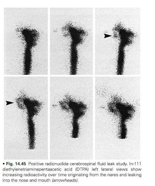
The site is most likely to be identified during a time when heavy leakage is occurring. Often, the patient position associated with the greatest leakage is reproduced during imaging. Imaging in the appropriate projection is important for identifying the site of leak; lateral and anterior imaging are used for rhinorrhea and posterior imaging for otorrhea. In cases in which no reason is known for low CSF pressure or when a leak around the lumbar region is suspected, additional views should be made of the lumbar region.

Scintigraphic studies show CSF leaks as an increasing accumulation of activity at the leak site.  

However, counting the pledgets is more sensitive than imaging for detect- ing CSF leaks. Pledgets are also helpful in determining the origin of the leak (anterior vs. posterior).


  • Follow me on Twitter @RutujaKote for more discussions about topics in Nuclear Medicine and Radiology.


Monday, December 5, 2022

Chronic Cholecystitis on Cholescintigraphy

  • Introduction: 
  • Recurrent episodes of right upper quadrant pain, usually in a middle- aged female, although occasionally in men and children, are suggestive of chronic cholecystitis. 
  • The clinical diagnosis is often confirmed by detection of gallstones on sonography. 
  • The standard therapy is cholecystectomy; gallbladder histopathology shows evidence of chronic inflammation. 
  • On occasion, a clinician suspecting that a patient’s pain is not due to cholecystitis and that the gallstones seen are incidental may refer the patient for cholescintigraphy and a GBEF. 
  • Although chronic cholecystitis classically shows delayed filling after 60 minutes, some cases will show normal gallbladder filling. 
  • When CCK is administered after gallbladder filling, patients with asymptomatic cholelithiasis will have normal gallbladder contraction, whereas patients with chronic cholecystitis have a poor gallbladder response.

Chronic Acalculous Gallbladder Disease

  • The acalculous form of chronic cholecystitis occurs in approximately 10% of patients with symptomatic chronic gallbladder disease. 
  • It is clinically and histopathologically indistinguishable from chronic calculous cholecystitis, except that there are no gallstones. 
  • This entity has been called by various names in the literature and by referring physicians, including gallbladder dyskinesia, gallbladder spasm, cystic duct syndrome, and functional gallbladder disease. 
  • Patients present with recurrent right upper quadrant biliary colic, have poor gallbladder contraction, and are usually cured with cholecystectomy.
  • Many investigations have found that sincalide cholescintigraphy can confirm the suspected clinical diagnosis of chronic acalculous gallbladder disease. 
  • A poor GBEF predicts postcholecystectomy symptomatic relief and histopathological evidence of chronic gallbladder inflammation; a normal GBEF excludes the disease. 
  • There is only one small (21 patients) randomized prospective study. In that study, 92% were cured with surgery. 
  • Sincalide cholescintigraphy should be performed on an outpatient basis after a clinical evaluation has excluded other diseases. 
  • It is best performed as an outpatient because acute illnesses and numerous therapeutic drugs can adversely affect gallbladder contraction and result in a false-positive study.

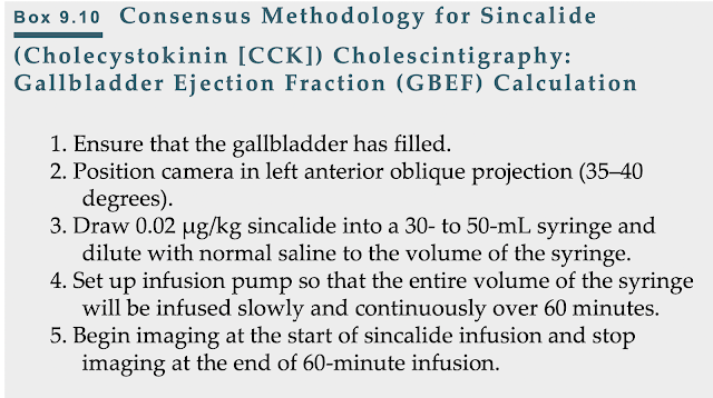
Sincalide Infusion Methodology

  • A consensus report published by expert gastroenterologists, surgeons, and nuclear medicine physicians recommended that the 60-minute infusion method should become the standard methodology.

  • Follow me on Twitter @RutujaKote for more discussions about topics in Nuclear Medicine and Radiology.
Friday, December 2, 2022

Acute Cholecystitis on Cholescintigraphy

  • The most frequent indication for cholescintigraphy is to confirm or exclude the diagnosis of acute cholecystitis. 
  • Patients present with colicky right upper quadrant abdominal pain, nausea, and vomiting. 
  • Physical examination often detects right upper quadrant tenderness. Laboratory studies show leukocytosis.
  • Series of sequential histopathological inflammatory changes occurs—first, venous and lymphatic obstruction, followed by edema of the gallbladder mucosa, then white blood cell infiltration, and, ultimately, hemorrhage, ulceration, necrosis, and if left untreated, gangrene, abscess, and perforation.

  • USG:
  • Most patients with acute cholecystitis have gallstones noted on sonography; however, the presence of stones is not specific for acute cholecystitis.
  • Asymptomatic gallstones are common and may be unrelated to the cause of the abdominal pain.
  • Thickening of the gallbladder wall and peri-cholecystic fluid occur with various acute and chronic diseases.
  • A more specific indicator of acute inflammation is intramural lucency. The sonographic Murphy sign (localized tenderness in the region of the gallbladder) is reported to have high accuracy in experienced hands; however, this finding is operator dependent and not always reliable. The combination of gallstones, intramural lucency, and the sonographic Murphy sign makes the diagnosis of acute cholecystitis likely.
  • Ultrasonography may reveal other factors causing the patient’s symptoms (e.g., common duct dilation due to biliary obstruction, pancreatic or liver tumors, renal stones, pulmonary consolidation).

  • Cholescintigraphy:
  • A major advantage of HIDA scintigraphy is that it demonstrates the pathophysiology of acute cholecystitis (i.e., nonfilling of the gallbladder secondary to cystic duct obstruction). 
  • No filling by 60 minutes after Tc-99m HIDA injection is abnormal; however, it is not, by itself, diagnostic of acute cholecystitis.
  • However, no filling on further delayed imaging at 3 to 4 hours or 30 minutes after morphine infusion is diagnostic of acute cholecystitis.
  • Delayed filling of the gallbladder (i.e., after 60 minutes) rules out acute cholecystitis. Common reasons for delayed gallbladder filling are chronic cholecystitis and hepatic dysfunction
  • Cholescintigraphy has high accuracy for the diagnosis of acute cholecystitis. The sensitivity (nonfilling of the gallbladder in those with the disease) is 95% to 98%, and the specificity (filling of the gallbladder in patients who do not have the disease) is > 90%.
  • Ensuring that patients have fasted for 3 to 4 hours before the study is critical. Those fasting > 24 hours or receiving hyperalimentation likely have a gallbladder full of viscous bile and should be administered sincalide before the study to empty the gallbladder. 

  • False Positive:
  • In these cases, false positives may still occur because of a poorly contracting gallbladder in response to CCK due to chronic cholecystitis. Patients with poor hepatic function have delayed uptake and clearance of the radiotracer and often delayed gallbladder filling. In these patients, delayed imaging for up to 24 hours may be necessary to confirm or exclude gallbladder filling; false positives may still occasionally occur.
  • Patients with chronic cholecystitis may have false-positive findings for acute cholecystitis (nonfilling of the gallbladder) caused by a fibrotic obstruction of the cystic duct or a functional obstruction caused by a gallbladder filled with viscous bile. Even if a patient has received sincalide before the study, a diseased gallbladder, whether acute or chronic, may not contract. 
  • Very ill hospitalized patients with a concurrent serious illness may also have false-positive scintigraphic results for acute cholecystitis. The reason for this is uncertain.

  • False Negative:
  • False-negative results (gallbladder filling in a patient with acute cholecystitis) are rare. One important cause to be avoided is misinterpretation of the cystic duct sign, specifically cystic duct dilation proximal to its obstruction, which might be misinterpreted as a gallbladderThe focal activity is typically smaller than a gallbladder and in a more medial position

  • If the gallbladder does not fill by 1 hour, either delayed imaging for up to 4 hours or morphine administration is indicated to confirm or exclude gallbladder filling. 

  • Morphine Administration:
  • The accuracy of morphine is similar to the delayed imaging method and is preferred whenever possible because it confirms or excludes the diagnosis by 30 minutes after administration. 
  • Morphine produces a functional partial common duct obstruction that cannot be differentiated by scintigraphy from a pathological partial common duct obstruction caused by stone or stricture. 
  • Thus, morphine should not be administered if scintigraphic findings show delayed clearance from the common duct and delayed transit into the small bowel. 
  • Delayed imaging is indicated for these patients. 
  • With cystic duct patency, the gallbladder begins to fill within 5 to 10 minutes after morphine infusion and is complete by 20 to 30 minutes. 
  • If no gallbladder filling is seen by the end of the 30-minute infusion, acute cholecystitis is confirmed.
  • Ancillary Findings:
  • Increased blood flow to the gallbladder fossa secondary to severe inflammation is seen in some patients. 
  • Increased hepatic uptake of HIDA tracer adjacent to the gallbladder fossa in patients with acute cholecystitis is called the rim sign and is seen in approximately 25% of patients with acute cholecystitis. 
  • The rim sign is more common than increased flow to the gallbladder fossa. Sometimes they occur together. 
  • The rim sign can usually be seen throughout the duration of the study but is best seen as the radiotracer clears from the uninvolved liver. 
  • It is caused by inflammation of the liver adjacent to the gallbladder fossa. 
  • With severe acute cholecystitis, inflammation may spread to the adjacent normal liver, which can result in increased blood flow to that region, increased radiotracer delivery, and thus increased Tc-99m HIDA hepatic extraction.
  • The importance of the rim sign is twofold. 
  • First, it is a very specific scintigraphic finding of acute cholecystitis. It increases interpretive confidence that nonfilling of the gallbladder is caused by acute cholecystitis (true positive) in a patient at increased risk for a false- positive study, for example, a sick hospitalized patient with concurrent serious illness. 
  • Second, the rim sign identifies patients with acute cholecystitis who have more severe disease and are at increased risk for complications (e.g., gangrene and perforation). 
  • Even without these complications, patients with the rim sign tend to be sicker and at a later stage of the pathophysiological spectrum of disease, with hemorrhage and necrosis rather than edema and leukocyte infiltration.

  • Follow me on Twitter @RutujaKote for more discussions about topics in Nuclear Medicine and Radiology.

Thursday, December 1, 2022

Cholescintigraphy: Normal Findings

 Normal Physiology of Liver:

  • Liver –secretes  600 mL of bile/ day  (0.4 mL/min)
  • 50%  enters  the  gallbladder (0.2  mL /min)
  • Normal of GB capacity-50 mL
  • Absorption of water –“concentration  function  of  the gallbladder”
  • Mean basal pressure 
    • Gallbladder lumen -10 cm of water
    • CBD -12 cm of water 
    • Sphincter of Oddi -15 cm of water 
  • Absorption of water through the wall  is one  of two main  factors  that  facilitates hepatic bile entry  into the gallbladder
  • Increase in  the  tone  of  the sphincter of Oddi
Gall Bladder Contraction and Emptying:
  • Nervous 
  • Endocrine 
  • Nervous 
  • Parasympathetic (vagus) -stimulate  contraction of  the  gallbladder
  • Sympathetic (celiac ganglion)-relaxation of  the  gallbladder  
  • Normal function of  the gallbladder -after its denervation
  • Role of the  sympathetic and parasympathetic nervous system – very minimal 
  • Primary regulator  of  gallbladder  emptying  is  the  hormone.

  • Cholecystokinin (CCK)
  • CCK-33 –by I cells of upper intestine- when food enters the duodenum
  • Acts through CCK (A & B)receptors  in  the  smooth muscle 
  • The biological action of the hormone  resides in its terminal eight  amino  acids
CCK
  • Contracts gallbladder
  • Relaxes the sphincter of Oddi 
  • Threshold for sphincter of Oddi relaxation –is much lower
  • May stay  relaxed  for  a  longer  period  of time
  • The  commercially  available hormone -CCK-8
  • Attach to  the  same  receptors  
  • Degree of  gallbladder emptying relates directly to the number of CCK receptors
Radiopharmaceuticals:
  • I 131 rose bengal
  • Suboptimal imaging characteristics  
  • Relatively high radiation dosimetry limited its clinical utility

  • Tc -99m-iminodiacetic acid (99mTc-IDA) 
  • Tc-99m-diosopropyl-IDA (DISIDA) or disofenin (Hepatolite)
  • Tc-99m-bromotriethyl-IDA (BrIDA) or mebrofenin (Choletec)
  • 99mTc-IDA – follows same hepatocyte uptake, transport and excretion pathways as bilirubin
  • Competitive inhibition
  • Radiotracer transport into hepatocyte by high capacity carrier mediated anion clearance mechanism
  • Tracer is transported into Bile canaliculi by an active membrane transport system similar to bilirubin
  • Mebrofenin –Rp of choice in jaundice.
  • Least bound to be involved by inhibition from bilirubin
  • Mebrofenin is useful even at bilirubin of 20 -30 mg/dl
Normal HIDA Findings:

  • Clearance of blood pool activity -5 min
  • CBD is usually seen by 20 min
  • Gall bladder – begins to fill by 10 min

In 90% normal individuals- GB is filled by 30-40 min

In 5% to7% normal individuals- GB is noted  late,  between 50 to 60 min

  • Gut- is usually visualized by 60 min


  • Follow me on Twitter @RutujaKote for more discussions about topics in Nuclear Medicine and Radiology.